Two patients walk into a clinic with nearly identical MRIs. One of them has been in debilitating pain for months. The other has no symptoms at all. Same findings on film. Completely different lives. If the MRI were the whole story, that should not be possible. But it is, and it happens more often than most patients know.
This is one of the most uncomfortable realities in spine care. The image and the person do not always match.
What the MRI Is Actually Showing You
An MRI is a structural snapshot. It shows the spine at rest, in a controlled position, at a single moment in time. It is excellent at identifying disc herniations, nerve compression, and degenerative changes. For certain diagnoses it is indispensable. But it captures the spine the way a photograph captures a runner mid-stride. It tells you something about that moment. It does not tell you how the whole race has gone.
What imaging does not show is how forces are moving through your spine. It does not show which segments are moving too much, which have lost motion entirely, or how your body has been compensating for years to keep you upright. Those compensation patterns are often where the real story lives, and they are invisible on a standard MRI.
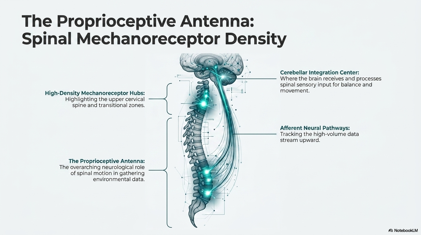
The Blueprint Every Spine Has
Every spine has a blueprint. There are fixed structural angles in the pelvis and the base of the neck that define what your spinal curves should ideally look like. Those angles do not change based on how you stand or how you feel on a given day. They are built into your bones.
What does change is how far your spine has drifted from that blueprint, and how your body has adapted to that drift.
The spine’s primary job is to keep your head balanced over your hips against gravity, using as little muscular effort as possible. When posture shifts away from the ideal, whether from injury, years of sitting, or gradual degeneration, the body responds with a predictable chain of compensations. The pelvis tilts. The mid-back rounds. The head shifts forward. Each adaptation keeps you functional but at a mechanical cost. More muscle work. More pressure on the discs. More stress on the joints over time.
A standard MRI captures that compensated state. It can make a spine look more normal than it actually is. What appears to be a lumbar disc problem on imaging may trace back to how the head is sitting over the pelvis. Treat only the disc and you have treated a symptom of a pattern, not the pattern itself.
What Gets Missed Without Biomechanical Assessment
When imaging is the only lens, the evaluation captures what has already broken down. It does not capture the mechanical forces that drove the breakdown or where those forces are actually originating.
A thorough biomechanical assessment looks at the spine as a connected mechanical system. Full-spine X-rays with specific measurements evaluate the whole architecture, not just the region that hurts. Motion X-rays, taken as the patient bends or captured in real time video, show which segments are moving abnormally and what the ligaments are doing under load. That information points to where the mechanical work actually needs to happen, not just where it hurts.
The spine is not a collection of separate parts. A problem that shows up in the lower back may be driven by mechanics originating elsewhere in the chain. Addressing only the painful region without understanding what is loading it is treating the result without touching the cause.
What This Means for Your Care
If you have been told your MRI looks normal but you are still in significant pain, that is not a sign that the pain is imaginary. It is a sign that the full picture has not been assembled yet.
If you have imaging findings that were treated without improvement, it is worth asking whether the mechanical driver of those findings was ever identified.
Getting the right diagnosis matters more than anything else that follows. Not a general idea of what is wrong, but a specific and accurate picture that includes how your spine is actually moving and loading. Two patients with the same diagnosis on paper can need completely different treatment plans. The image alone cannot tell you which one you are.
The Evidence on Asymptomatic MRI Findings
The foundational studies on MRI findings in asymptomatic subjects were published in the early 1990s and have since been replicated many times. Boden and colleagues (1990) examined the MRI scans of 67 individuals with no history of back pain, sciatica, or neurogenic claudication. They found that 20 percent of subjects under 60 years of age had a herniated nucleus pulposus on MRI, and 57 percent of subjects over 60 had a significant abnormality, including herniation, stenosis, or degenerative changes. None of these individuals had any symptoms. The study was published in the Journal of Bone and Joint Surgery and remains one of the most cited papers in spine care literature.1
Jensen and colleagues (1994) published a similar study in the New England Journal of Medicine examining 98 asymptomatic subjects with lumbar MRI. They found disc bulges in 52 percent, disc protrusions in 27 percent, and facet degeneration in 8 percent. Only 36 percent of subjects had a completely normal scan. The study’s conclusion was direct: “The discovery by MRI of bulges or protrusions in people with low back pain may frequently be coincidental.”2 This was not a fringe finding. It was published in the highest-impact medical journal in the world and confirmed what clinicians who examined both symptomatic and asymptomatic patients had been observing for years.
A 2015 systematic review by Brinjikji and colleagues, published in American Journal of Neuroradiology, synthesized imaging findings from 3,110 asymptomatic individuals across 33 studies. The prevalence of disc degeneration in asymptomatic subjects increased from 37 percent at age 20 to 96 percent at age 80. Disc bulge prevalence ranged from 30 percent at age 20 to 84 percent at age 80. The authors concluded that many imaging findings commonly referred to as degenerative should be considered normal aging phenomena when found incidentally in asymptomatic patients.3
References
- Boden SD, Davis DO, Dina TS, Patronas NJ, Wiesel SW. Abnormal magnetic-resonance scans of the lumbar spine in asymptomatic subjects. J Bone Joint Surg Am. 1990;72(3):403-408.
- Jensen MC, Brant-Zawadzki MN, Obuchowski N, Modic MT, Malkasian D, Ross JS. Magnetic resonance imaging of the lumbar spine in people without back pain. N Engl J Med. 1994;331(2):69-73.
- Brinjikji W, Luetmer PH, Comstock B, et al. Systematic literature review of imaging features of spinal degeneration in asymptomatic populations. AJNR Am J Neuroradiol. 2015;36(4):811-816.
- Wiesel SW, Tsourmas N, Feffer HL, Citrin CM, Patronas N. A study of computer-assisted tomography. I. The incidence of positive CAT scans in an asymptomatic group of patients. Spine. 1984;9(6):549-551.
Ready for a Complete Biomechanical Evaluation?
Schedule with Dr. McClean at McClean Chiropractic in Provo, Utah. Fellowship-trained. Biomechanics-focused. The most advanced spine assessment in Utah County.