Digital Motion X-Ray (DMX) in Provo, Utah
See the hidden ligament injuries and spinal instability that static X-rays. And even MRI. Routinely miss. Motion Works DMX digital motion X-ray is performed on-site at McClean Chiropractic and read by our fellowship-trained doctors.
A "normal" X-ray doesn't mean nothing is wrong
Most patients who walk into an emergency room or urgent care after a car accident leave with a familiar message: “Your X-rays look fine. Nothing is broken.” And that’s technically true. But a static X-ray is a single frozen snapshot of your spine at rest. It cannot show how your vertebrae actually move relative to each other. Which is exactly where most post-crash and chronic-pain injuries live.
Ligaments, for example, are invisible on a standard X-ray. So are the motion-dependent pinches, slips, and instability patterns that cause most lingering neck pain, headaches, and radicular symptoms after a whiplash event. A patient can have a catastrophically injured alar or transverse ligament and walk out of the ER with a clean bill of health. Only to struggle with dizziness, cervicogenic headaches, and neck pain for years because no one ever looked.
That’s the diagnostic gap motion X-ray was built to close.
-
Ligament laxity and instability. Injured ligaments don’t show up on static imaging
-
Motion-dependent nerve compression. A pinch that only happens when you turn your head
-
Facet joint dysfunction. Joints that lock, gap, or slip during movement
-
Post-whiplash cervical instability. The most under-diagnosed cause of chronic post-crash symptoms
-
Weight-bearing disc behavior. A disc that looks fine lying in an MRI scanner can herniate when loaded
The Blueprint of Motion
How motion X-ray fluoroscopy turns the spine’s movement into clinical data — and why fellowship-trained chiropractors are uniquely qualified to read it.
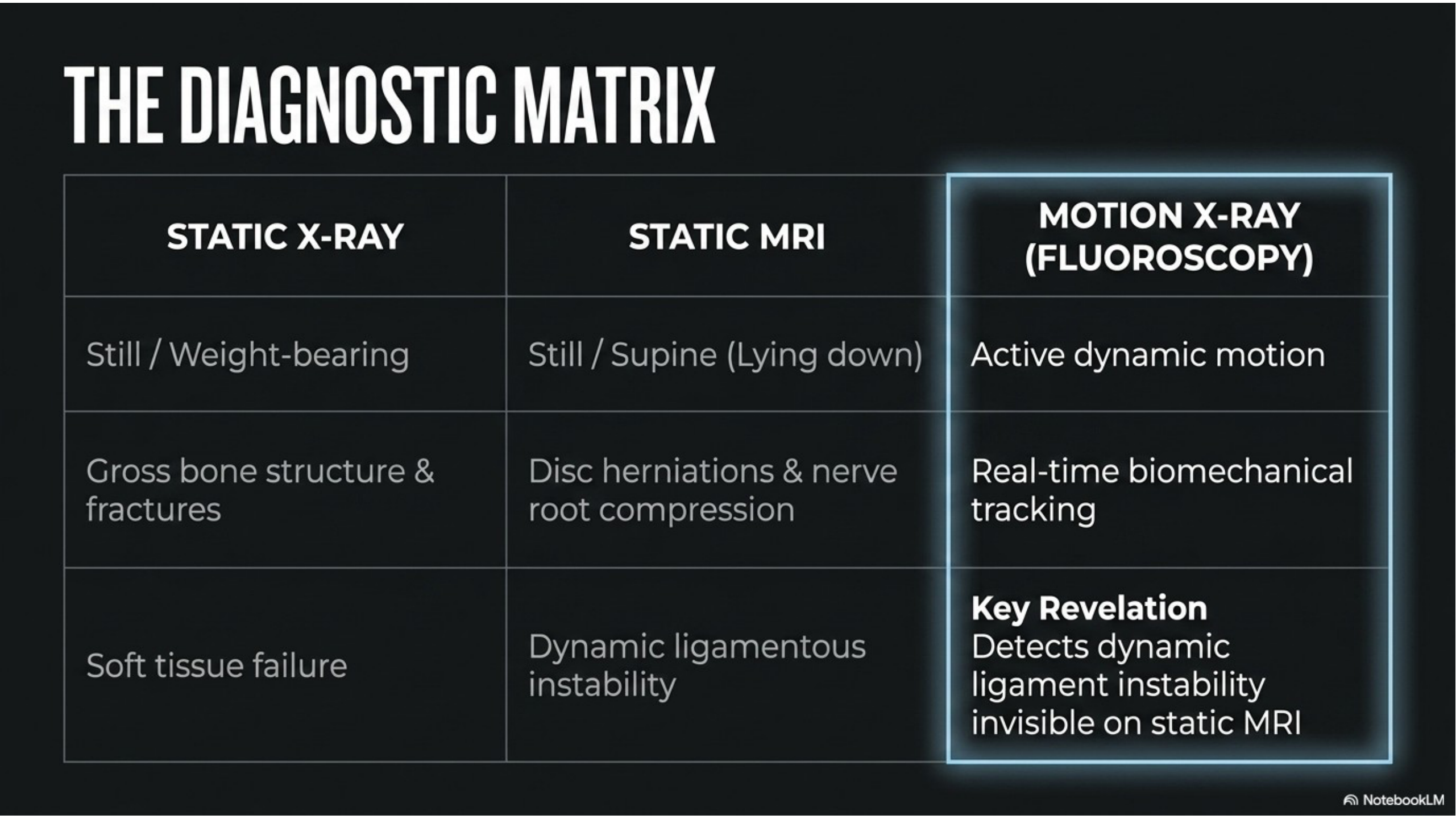
Static vs Motion Imaging
Motion X-ray uniquely detects dynamic ligament instability — invisible on every form of static imaging.
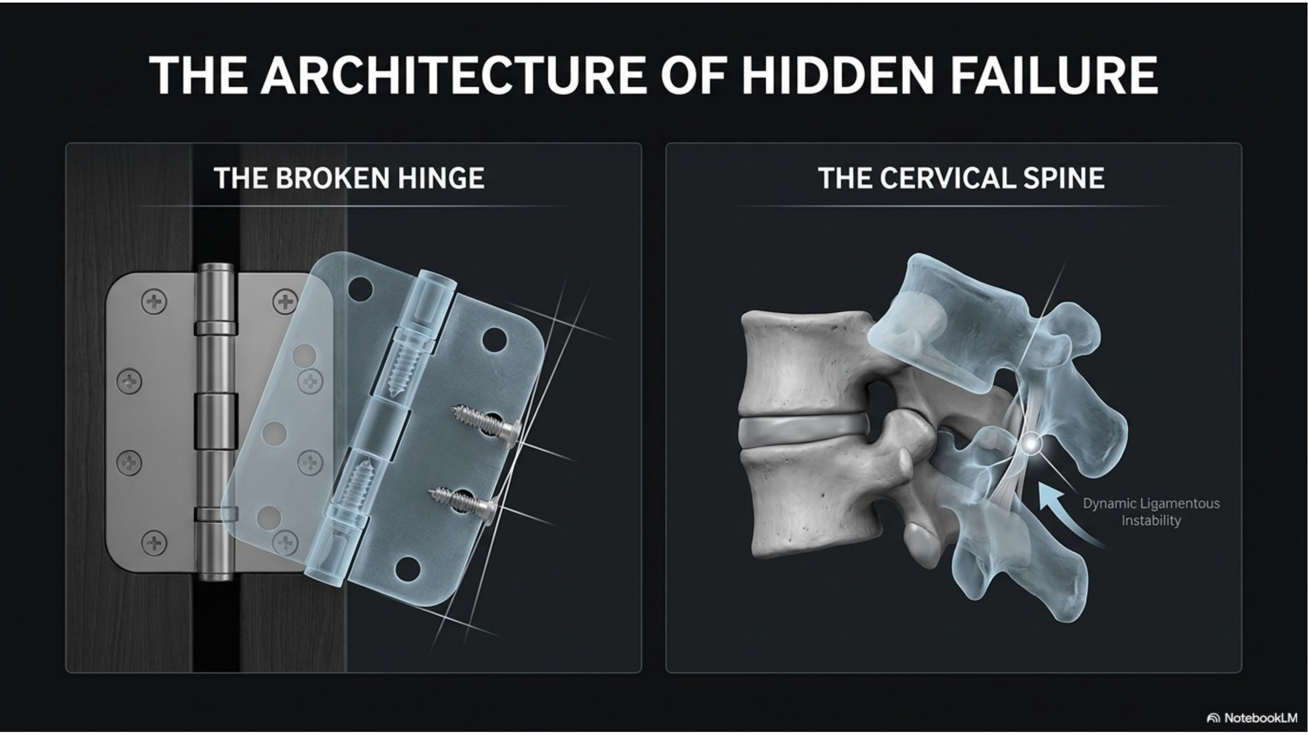
Hidden Mechanical Failure
Instability only reveals itself under load and in motion — exactly what static imaging cannot capture.
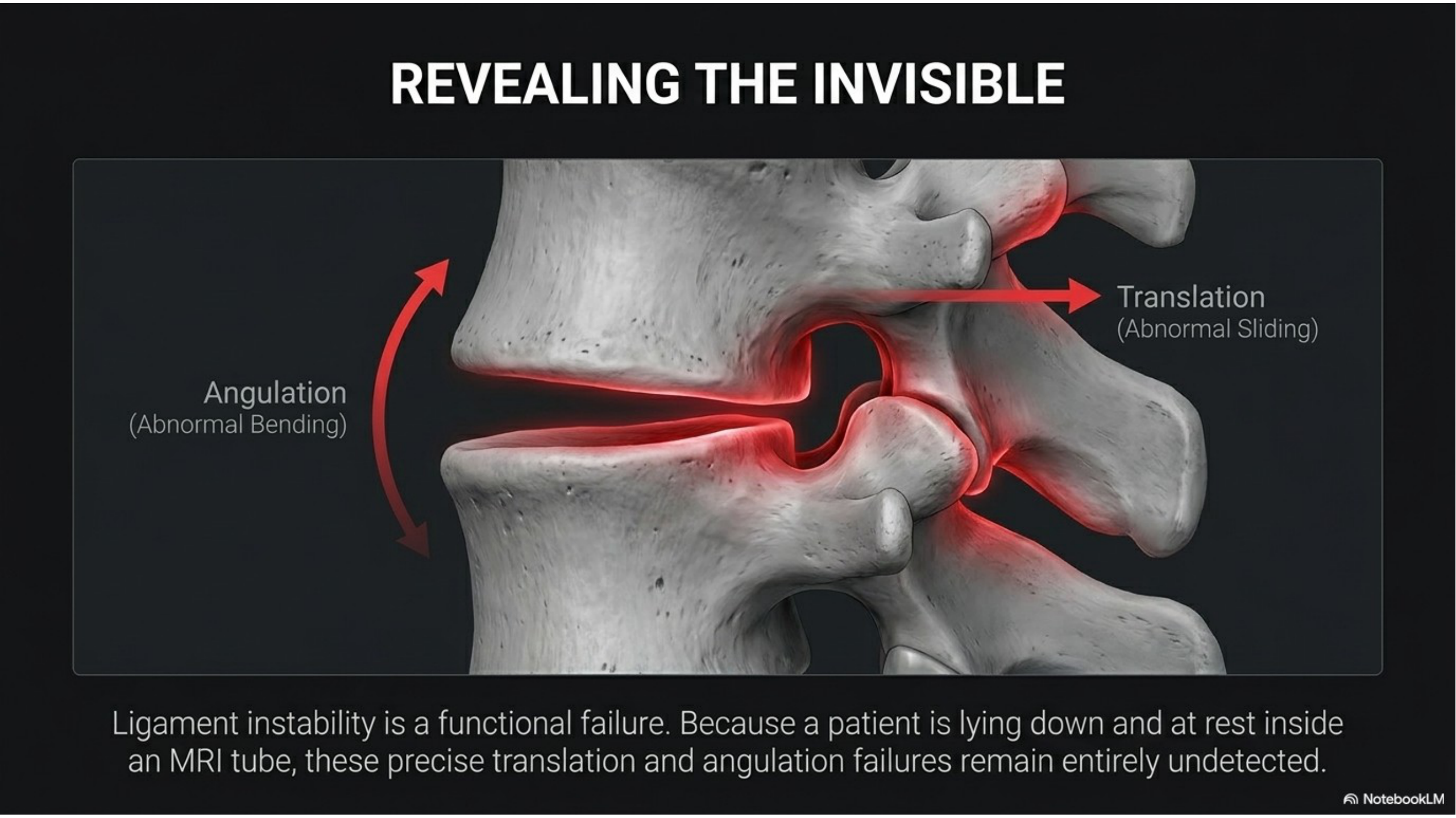
Revealing the Invisible
Abnormal angulation and translation become visible in real-time — pinpointing the exact segment that fails under load.
What is digital motion X-ray (DMX)?
Digital motion X-ray. DMX. Is fluoroscopic imaging captured as a continuous video while you actively move your spine. Instead of one static image, we record a video of the bones of your neck or low back while you flex, extend, side-bend, and rotate. Each frame can be analyzed individually, and AI-assisted measurement tools let us quantify exactly how each vertebra moves relative to its neighbors.
We perform the scan on-site with our DMX system. The same class of imaging technology used in medico-legal cases across the country.
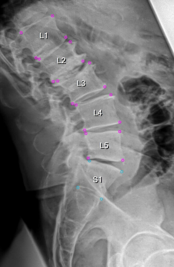
What motion X-ray reveals that static imaging can't
-
Cervical ligament injury. Alar, transverse, and anterior/posterior longitudinal ligament laxity
-
Atlanto-axial and atlanto-occipital instability. Often the hidden cause of chronic headaches and vertigo
-
Segmental hypermobility and hypomobility. Vertebrae moving too much or not at all
-
Flexion-extension aberrations. Paradoxical or stepped motion patterns after whiplash
-
Facet joint laxity and subluxation. Joints that open or slip under load
-
Quantified range-of-motion deficits. Hard numbers you can use for insurance, attorneys, and specialists
-
Medico-legal documentation. Objective imaging findings that can support personal injury, workers’ comp, and disability cases
Upper neck instability: what DMX can show
Some people with ongoing headaches, dizziness, a “heavy head” feeling, or neck symptoms that get worse when they’re upright have been told their MRI is normal. An MRI is usually taken lying down, with the head still. Instability in the upper neck, where the skull meets the top two vertebrae, may only show up when you’re upright and moving.
On a cervical DMX study, our doctors look for signs of upper-neck instability, including:
-
Abnormal sideways shifting of the first vertebra (C1) on the second (C2)
-
Widening of the gap between C1 and the dens (the peg of C2) when you bend forward
-
Excess movement between the skull and C1
-
Abnormal rotation patterns when you turn your head
-
Too much forward or backward sliding at each level as you bend and extend
DMX doesn’t diagnose craniocervical instability (CCI) on its own. What it gives you is upright motion imaging of your upper neck, read by a fellowship-trained doctor, with findings you can take to the specialists managing your care.
When should you consider motion X-ray imaging?
Motion X-ray is not for every patient. It’s a targeted diagnostic tool for situations where static imaging has failed to explain the symptoms, or where dynamic spinal behavior is clinically relevant. We’ll decide with you during your consultation whether a motion study is medically indicated.
-
After a car accident. Even a low-speed or "minor" collision. Ligament injuries do not scale with vehicle damage
-
Persistent neck or back pain that hasn’t resolved after weeks or months of treatment elsewhere
-
Post-concussion symptoms, chronic headaches, dizziness, or vertigo. Particularly when imaging has been "normal"
-
Your static X-rays or MRI came back clean but you still hurt. The next step is often dynamic imaging
-
You’re being evaluated for spine surgery. Understanding actual motion matters before any irreversible decision
-
Personal injury, workers’ comp, or attorney-referred cases. Motion imaging creates objective documentation
Why we perform motion X-ray on-site
Many clinics that offer motion imaging send patients to a separate facility. We have DMX on-site at our Provo clinic, so the entire diagnostic pathway happens in one building:
-
DMX on-site. No referral, no driving, no week-long wait
-
Fellowship-trained reading. Studies are read by our doctors, all three of whom hold the Fellowship in Spinal Biomechanics and Trauma (FSBT)
-
Custom cervical and lumbar protocols. Including flexion-extension, lateral bending, rotation
-
AI-assisted quantitative measurement. measured findings, not just “it looks loose”
-
Correlation with your MRI and physical exam. We read the whole picture, not just one study
Fellowship-Trained to Read Motion X-Ray
Motion X-ray fluoroscopy is a high-tech diagnostic tool — but the imaging is only as good as the doctor reading it. All three McClean doctors hold the FSBT (Fellow of Spinal Biomechanics & Trauma) credential.
That’s fellowship-level training in motion X-ray interpretation, MRI correlation, and AI-assisted analysis — a level of expertise most chiropractors never pursue.

What patients say about our diagnostic process

“I had tingling and numbness in one arm and back. Dr. Matt did X-rays and ordered an MRI, and found an impinged nerve. He carefully adjusted and his staff were very methodical about treatment. Within a three-week period I was totally back to normal. The business is operated like a well-oiled machine.”
Bobbi K.
“I was in Utah on vacation with serious radicular pain down my left leg. I called Monday morning before they opened and was seen right away. Dr. Matt did an X-ray, adjusted me, and scheduled an MRI for later that evening. By Tuesday morning I knew I had a posterolateral disc herniation at L5/S1. Everyone. Receptionist, assistants, doctor. Was A+ top notch.”
Johnny K.
Motion X-Ray. Frequently Asked Questions
Is motion X-ray safe?
Motion X-ray uses fluoroscopy, which exposes you to more radiation than a single static X-ray but substantially less than a CT scan. For patients with a documented injury or unresolved symptoms, the diagnostic value far outweighs the radiation exposure. Please tell us before any imaging if you are or might be pregnant.
Does insurance cover motion X-ray?
Most auto insurance (PIP and third-party), personal-injury attorney liens, and workers’ compensation cases cover motion X-ray when it is medically indicated. Medicare and major health insurance coverage varies by plan. Call our office at 801-373-1035 and we’ll verify your benefits before you come in.
How long does a motion X-ray appointment take?
The imaging itself is quick. When you schedule, we’ll let you know how much time to plan for.
Motion X-ray vs. MRI. Which one do I actually need?
They show completely different things. MRI is the best test for soft tissue. Discs, nerves, spinal cord, edema. Motion X-ray is the best way to see dynamic instability and ligament injury. If you’ve had whiplash, persistent post-crash symptoms, or your static imaging was “normal” but you still hurt, motion X-ray is usually the missing piece. Many patients benefit from both. And we also offer a Free MRI Review if you’ve already had an MRI.
What if my static X-rays from the ER came back normal?
That’s actually the most common reason patients come to us for motion imaging. Static X-rays rule out fractures and gross bone disease. They do a good job of that. But they cannot show ligament injury, instability, or motion-dependent nerve compression. A “normal” static X-ray after a car accident doesn’t mean nothing is wrong; it means the bones weren’t broken.
Do I need a referral from another doctor?
No. You can self-refer. We’ll determine during your consultation whether motion imaging is medically indicated based on your history and physical exam. If your attorney, primary care doctor, or a specialist has already requested the study, we can perform it and share the results with them.
Can you read motion X-rays performed at another facility?
Yes. If you already have an MRI from another facility, you can send it through our Free MRI Review page. For other outside studies, call us to ask.
Can DMX show craniocervical instability (CCI)?
It can show signs of it. A cervical DMX study records your upper neck moving while you are upright, which a standard MRI does not, and our doctors review it for abnormal motion between the skull, C1 and C2. A CCI diagnosis also depends on your history, exam and other imaging, so we explain what the study shows and you can share it with your other doctors.
Ready to see what's really going on?
McClean Chiropractic is one of the only chiropractic clinics in Utah County with on-site digital motion X-ray. Read by our fellowship-trained doctors. Call us and we’ll walk you through whether motion imaging is appropriate for your situation.
More From Our Spine Education Library
Articles from our fellowship-trained doctors on the topics above: